Home » News » Use 3D Histopathology to Improve Experimental Designs
Use 3D Histopathology to Improve Experimental Designs
Posted on Jun 06, 2022

3D IHC Doesn't Rely on Reconstruction of 2D FFPE Thin-section Slides
Your research will benefit from 3D histopathology whether you perform basic research at a Children's Hospital to better understand the underlying factors of a pediatric disease or you're in a promising biotech working on a novel therapeutic agent.
Life in three dimensions is more fulfilling. Imagine your family's summer vacation, stuck in 2D. It would be lackluster. Every family member would turn into the moping teenager, "Mom, why do we have to go to the flatland again?" Life is 3D. Biology is 3D. Your research applications should reflect that.
Leverage ClearLight for your Preclinical Research
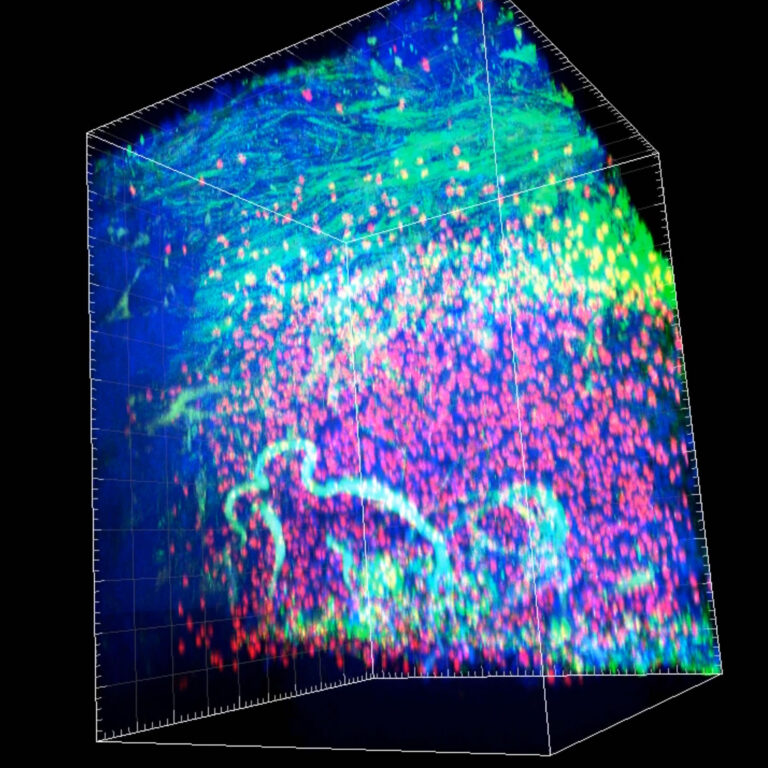
Preclinical researchers in academic and commercial environments leverage our expertise with CLARITY tissue clearing, thick tissue immunostaining, and Tru3D® tissue analysis to gain a better understanding of the tissue microenvironment. The CLARITY protocol enables researchers to work with larger tissue volumes to maintain tissue integrity and minimize the tissue/sample destruction associated with 2D thin section FFPE.
Examples of How ClearLight Bio's 3D IHC Can Empower Researchers:
- Understand the spatial relationships in the tissue microenvironment
- See where apoptosis and/or necrosis may be occurring
- Verify drug delivery to the intended tissue region
- Trace vasculature through the tissue
- Draw insightful conclusions leading to new and better questions
Push Your Preclinical Research Into 3D Territory
Are You Ready to Begin Your 3D Histopathology Journey?
Contact Us to Start a Pilot Project
The ClearLight Difference
- Analyzing 3D tissue is more biologically representative analysis of drug mechanism of action
- Deep expertise and intellectual property for CLARITY Tissue Clearing
- Experts at thick tissue antibody staining using a library of optimized antibodies
- Tru3D Tissue Analysis allows pathologists and researchers to see more biology than is currently possible with traditional 2D FFPE thin section methods
- Proprietary AI software to analyze 3D images (under development)
- CLARITY allows a tissue sample to be rendered optically clear, retain its three-dimensional integrity and allows for tissue staining and re-interrogation multiple times for most samples.
Our step-by-step approach maximized your insights while minimizing your risks.
- Each lab services engagement begins with a pilot project
- Our approach minimizes risk for you and your lab
- We process a small number of samples using our 3D IHC technology
- At the end you receive a summary report and data
- We review your results together and discuss potential next steps


