From 2D to 3D Tissue Imaging
Beyond Slices and Slides.
Biology isn’t 2D and your research shouldn’t be either.

Is Your Research Missing the Bigger Picture?
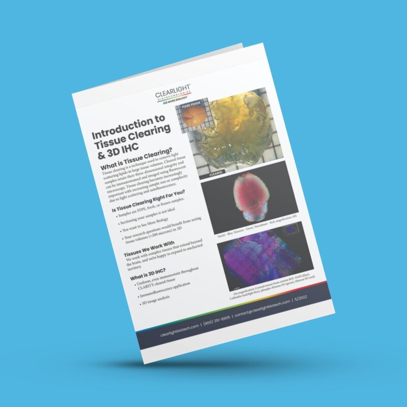
If your research relies on 2D FFPE thin sections (~5-10 microns) you are missing the bigger picture. Vasculature and structures in the tissue microenvironment don’t always follow predictable pathways. Increasing tissue thickness can be a challenge, too. As tissue thickness increases, light scattering and auto-fluorescence become issues. These are problems easily overcome with 3D IHC.
Beyond Thin Sections
Move beyond slices and slides. The future is 3D IHC, because Biology isn’t 2D and your research shouldn’t be either.
“The images we received from ClearLight led to us revising our direction and cutting our losses on an approach that wasn’t working. In the end we estimate that insight saved us at least 1000 hours in staff time that could then be more profitably utilized.”
Why be Limited?
When you need 3D answers but are working with 2D slides; it’s time for 3D IHC.
“We had tried several other methods prior to working with ClearLight, but the results were inconclusive and unsatisfactory. The video from the 3D IHC finally got us the confirmation that our experimental therapeutic may be working.”
Move Beyond Slices and Slides
Stop Using 2D Solutions to Solve 3D Problems
Reduce Uncertainty of Slices and Slides
Maximize Insights. Minimize Risk.
Each lab services engagement begins with a pilot project.
- Our approach minimizes risk for you and your lab.
- We process a small number of samples using our 3D IHC technology.
- At the end you receive a summary report and data.
- We review your results together and discuss potential next steps.
See the results you can get from 3D IHC, while minimizing cost and time.


