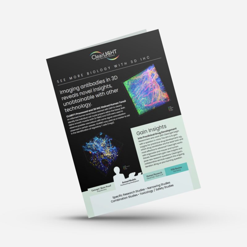
Preclinical Drug Development
Deserves an Early Advantage
Work with ClearLight to advance a better drug candidate.

Preclinical Drug Development
Long before your team will experience commercial success with your brand name drug, preclinical drug development activities take center stage. By the time you submit your investigational new drug (IND) application you’ll want to retire as many risks as possible and have confidence that your experimental drug will have its intended effects on the intended to treat population (ITT). Getting a strong drug candidate to the IND phase is a major step on your path to commercial success.
ClearLight in the Preclinical Drug Development Pipeline

“The juxtaposition of new insights into human biology, coupled with the application of new tools and advanced technologies, has the potential to revolutionize our business more in the next decade or two than in the past five!”
- John C. Lechleiter, PhD, Chairman, President, & Ceo, Eli Lilly & Company
Give your Preclinical Drug Development Program an Early Advantage
The majority of new drug candidates fail before making it to Phase I Clinical Trials. Preclinical studies must demonstrate the biological activity of the drug against the targeted disease. The drug candidate must also be evaluated for safety. These studies can take several years to complete.
Once a potential treatment shows promise to advance to Clinical Trials it still has a large fallout. Most potential drugs never make it to the medicine cabinet. Could improved earlier studies increase the chances for advancement? At ClearLight we think the answer is yes. We believe preclinical researchers should understand how a given therapy works in tissue before getting to the high stakes world of clinical trials. ClearLight empowers researchers to See More Biology™ within the tissue microenvironment.
Leverage ClearLight for your Preclinical Drug Development
Preclinical drug developers in academic and commercial environments work with ClearLight to gain an early advantage in their quest to advance effective therapeutics. Preclinical researchers leverage our expertise with CLARITY tissue clearing, thick tissue immunostaining, and Tru3D® tissue analysis to gain a better understanding of the tissue microenvironment.
The CLARITY protocol enables researchers to work with larger tissue volumes to maintain tissue integrity and minimize the potential cell destruction associated with 2D thin section FFPE sample preparation.
Researchers who work with ClearLight,
See More Biology™
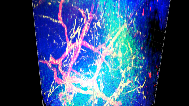
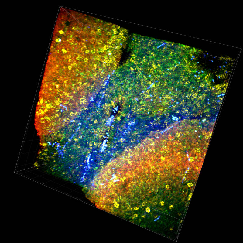
Imaging biomarkers in three dimensions reveals novel insights unobtainable with 2D FFPE and other technology:
- Understand the spatial relationships in the tissue microenvironment
- See where apoptosis and/or necrosis may be occurring
- Verify drug delivery to the intended tissue region
- Trace vasculature through the tissue
- Draw insightful conclusions leading to new and better questions
CLARITY Beyond the Brain
Chances are, ClearLight has worked with your research tissue type. ClearLight has evaluated numerous normal and diseased tissues. Our scientists have taken CLARITY beyond the brain to include organoids, eye, lymph nodes, skin, calcified tissues (bone and teeth), kidney, breast, colon, lung, pancreas, liver, tonsil, and other human and animal tissues.
ClearLight pushes the envelope of tissue clearing with continued innovation of its products and services.
The ClearLight Difference
- More biologically representative analysis of drug mechanism of action by analyzing 3D tissue
- Deep expertise and intellectual property for CLARITY Tissue Clearing
- Experts at thick tissue antibody staining using a library of optimized antibodies
- Tru3D Tissue Analysis allows pathologists and drug researchers to see more biology than is currently possible with traditional 2D FFPE thin section methods
- CLARITY-based tissue clearing services offer 10X more contiguous and more meaningful data as compared to current gold standard 2D FFPE thin-sections
- Proprietary AI software to analyze 3D images (under development)
- CLARITY allows a tissue sample to be rendered optically clear, retain its three-dimensional integrity and allows for tissue staining and re-interrogation multiple times.
Why Collaborate with ClearLight Biotechnologies?
ClearLight is a collaborative research organization. Unlike traditional contract research organizations that want to put you in a box, we listen and co-design an approach that answers your research question. Not all research follows a straight path. As our service engagement unfolds, new questions often arise.
“Sometimes you don’t get the result you’re hoping for. But data, whether affirming or unexpected, always leads to greater understanding through research.”
- Dr. Sharla White, VP of Research and Development
Reduce Risk and Increase Velocity Toward an IND
- Avoid going down a blind alley
- Confront and navigate around unexpected results
- Eliminate blind spots
- Identify needed modifications early
Schedule an Exploratory Call
See if CLARITY and 3D IHC are a good fit for your preclinical drug development research.
Introduction to CLARITY and 3D IHC
Our Mission:
ClearLight empowers researchers to see more biology within the tissue microenvironment. We are developing technologies to improve diagnostic, prognostic, and predictive treatment of disease so we can all live healthier lives.
Learn More About Our Services & Capabilities
Explore ClearLight Lab Services
Lab Services include CLARITY tissue clearing, 3D immunostaining, and Tru3D tissue analysis.
Implantable Device Case Study
See how ClearLight helped a biomedical innovator design a better implantable device for drug delivery.
Blog: Power of Retrospective Studies
Work with ClearLight to perform a retrospective study and get more value from your precious tissue samples.


