Immunohistochemistry
3D IHC Multiplexing
with CLARITY Tissue Clearing and Tru3D Imaging
3D Immunohistochemistry
Modern medicine is filled with technology, specialization, and science that builds upon earlier work. Like other science and technology, immunohistochemistry (IHC) has now been assimilated into the general body of medical knowledge. IHC is now an indispensable tool for the diagnosis, prognosis and treatment of disease as well as being a foundational tool for biomedical research and development around the world. That wasn’t always the case. IHC was built on the shoulders of giants, one scientific giant in particular, Dr. Albert Coons.
When You’re Ready for 3D Immunohistochemistry
ClearLight offers antibody-based immunostaining or labeling for many nucleic acids (RNA/DNA) to highlight specific structures in the CLARITY tissue cleared sample. Multiplexed immunofluorescence staining enables advanced Tru3D™ tissue analysis.
3D IHC Inspiration
The very foundation of Immunohistochemistry hearkens back to 1939 when Dr. Albert Coons was on vacation in Berlin, Germany. Dr. Coons was pondering ways to locate and identify antibodies as part of his study of rheumatic fever. On that fateful trip he gained the insight that he could put a visible label on an antibody molecule. This would allow targeted proteins to be seen within a diseased or normal tissue using a light microscope. He envisioned they would become brightly fluorescent. The concept was eventually born out and the idea took hold. Other researchers in laboratories around the world began using this method and the technique of immunofluorescence flourished.
3D Immunohistochemistry Video
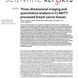
ClearLight Published Papers Related to Immunohistochemistry
Services
No. ClearLight Biotechnologies provides study-related services to sponsors of drug research programs early in the drug development cycle. Our contract research and development services are for Research Use Only (RUO).
Technology
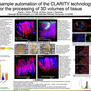
We use CLARITY tissue clearing to render opaque tissues primed for deep tissue imaging. Once tissues are lipid-cleared they can be immunostained and imaged in 3D. We have found the order clear-stain-image matters. See our Tissue Clearing Comparison
Imaging biomarkers in 3D reveals novel insights not available with other technologies. Biological tissues are complex and intrinsically 3-dimensional. We work with researchers who want to view spatial relationships in X, Y, and Z dimensions to gain new understanding of the tissue microenvironment.
Staining can be performed on the tissue sample without interference from the hydrogel matrix. With CLARITY tissue clearing, the sample is processed via Multiplex IHC staining, allowing the characteristics of the tumor microenvironment to be fully analyzed in three dimensions.
Unlock New Research Possibilities
When you choose 3D IHC for your research it is important to find the right antibodies and the right protocol that will address the research questions you have. To achieve the best results for clearing, antibody-based immunostaining, and imaging we encourage you to leverage ClearLight’s expertise and intellectual property. Get in touch with us when your team is ready to unlock new possibilities for your research.
Immunohistochemistry Related Services
CLARITY Tissue Clearing Primer
Need to know more about CLARITY Tissue Clearing? Visit the CLARITY tissue clearing page to learn the background and specific steps in the ClearLight CLARITY Tissue Clearing Process. Download the CLARITY Tissue Clearing Infographic.
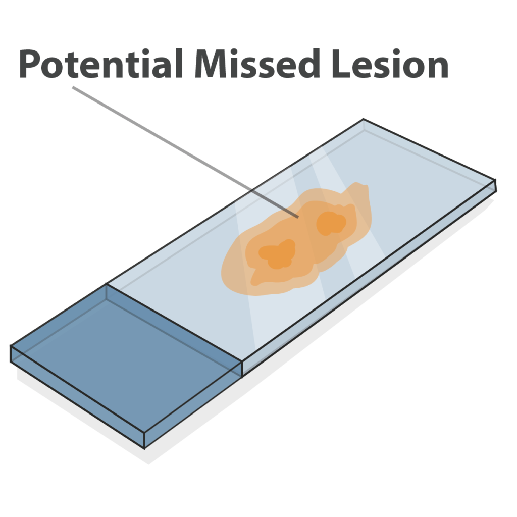
Compare FFPE to CLARITY + Tru3D
Imagine not being able to image antibodies in 3D. Many researchers are stuck in that world. Cancer doesn’t limit itself to two dimensions. Why should you and your research. See the difference between 2D FFPE and CLARITY + Tru3D.

Optimized Antibodies for CLARITY
Multiplexed IHC enables definitive antibody expression that is clear and easy to visualize. ClearLight has many optimized antibodies ready to use with CLARITY and 3D IHC to move your research forward. A feasibility study for new antibodies is available.
CLARITY Processed and IHC Stained Tissue Examples

CLARITY Processed and 3D IHC Stained Breast Tissue
Innovation Follows Discovery
Most science and technology breakthroughs are followed by additional innovations after the initial discovery. Innovators see new connections and forge new patterns. Hunches become hypotheses. Experiments are designed and conducted. Insights are gained, papers published and further conversations stoked resulting in a new cycle of discoveries.
CLARITY is one such discovery. Because most tissues are not transparent, and imaging processes are limited by light scattering; imaging deeply within tissues historically has been problematic. But with the complete removal of intracellular lipids during tissue clearing, immunostaining depth and quality improves. And because a CLARITY tissue-cleared sample remains intact, multiple rounds of fluorescent labeling could be possible. Whole tissue samples become reusable. That is helpful because who among us has only one scientific question, only one biomarker of interest?

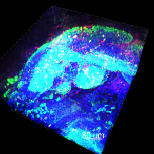
Classified and Segmented Tonsil Cells
Immunohistochemistry Staining
Next Generation Image Analysis
Another innovation that Dr. Coons could only have dreamed of are the advances in computing, specifically in machine learning, artificial intelligence, and image analysis. It is now possible to gather immunofluorescent data from multiple rounds of tissue staining and then individually segment and classify cells. A world of data is useless unless you can visualize it, process it, interpret, and understand complex relationships.
Biological tissues are complex and intrinsically three-dimensional. When researchers can view spatial data in X, Y, and Z dimensions the understanding of the tissue microenvironment can spark new clinical discoveries. This is a capability we are developing at ClearLight Biotechnologies. Imagine this capability for your next experiment.

CLARITY Processed and 3D IHC Stained Breast Tissue
What Will You Discover?
See More Biology with Multiplex IHC in 3D
Medicine and science continue to evolve. CLARITY tissue-cleared samples combined with 3D immunohistochemistry and Tru3D Alpha* tissue analysis gives researchers more data and more ways to envision that data.
These innovations empower scientific and medical discovery. Researchers can conceive of complex experiments, make new discoveries and invent better biomarkers that are diagnostic, prognostic and predictive of disease response.
Whether your revolution is in cancer research, and more specifically immunooncology research and development, neurodegenerative disease or infectious diseases, you owe it to yourself to work with our technology and the scientific team that is developing it.
