Tissue Clearing Applications
Breast Cancer
Learn More About Tissue Clearing
- Tissue Clearing Methods Compared
- CLARITY Tissue Clearing Primer
- CLARITY Breast Cancer Study
- Moving Beyond Slices and Slides
Supporting Your Research Application
Tissue Clearing Breast Cancer
ClearLight has performed tissue clearing on breast cancer and has extensive expertise in three-dimensional imaging and quantitative analysis of CLARITY processed breast cancer tissues.
Video of breast cancer tissue that was cleared using CLARITY protocol then immunostained with antibodies against pan-CK (green), Ki67 (yellow), CD3 (red), and a DAPI nuclear counterstain (blue).
Comparison of 3D Volume Merged View and 2D Optical Slice Section
Demonstrates Staining Uniformity and Visible Cell Classification
Tissue Clearing Breast Cancer
Biopsy Samples
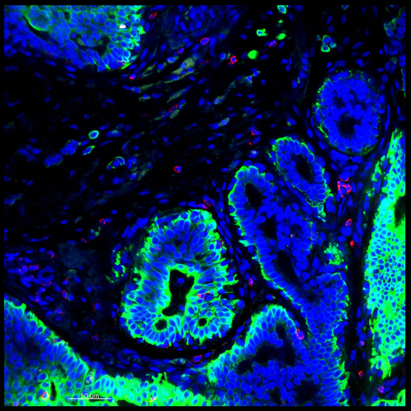
The CLARITY process was performed on previously fixed breast cancer core needle biopsy samples. Imaging on a confocal microscope (25x) clearly demonstrates how the tumor microenvironment is preserved. The 3D volume is shown in a merged view (left), with a 2D optical slice section (right) from the same volume that demonstrates the staining uniformity and visible cell classification. Subcellular staining was observed throughout these thick tissue sections. The breast tissue was immunostained with pan-CK (green), Ki67 (yellow), CD3 (red), and a DAPI nuclear counterstain (blue).
Award Winning Breast Cancer Study
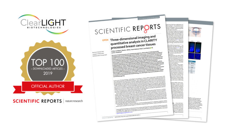
You might also be interested in this study where we determined the feasibility of a CLARITY tissue-processing approach to analyze biopsies from breast cancer patients. It was awarded Top 100 downloaded cancer papers on Scientific Reports.
“Three-dimensional Imaging and Quantitative Analysis in CLARITY Processed Breast Cancer Tissues,” awarded as one of the top 100 downloaded cancer papers.
Try tissue clearing on your own.
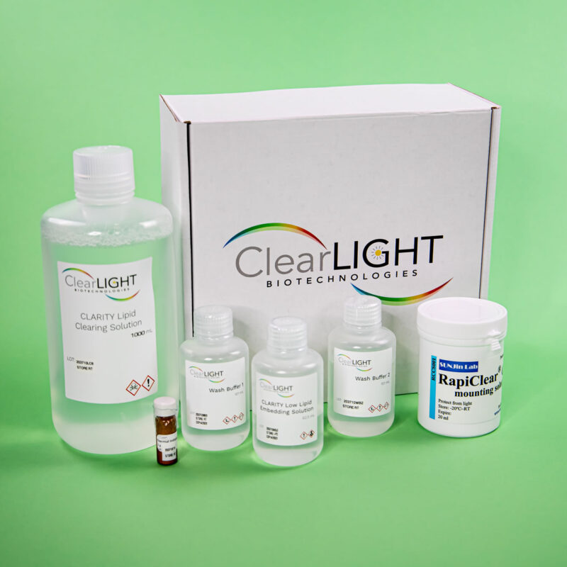
If you want to apply CLARITY to the tissues highlighted on this page, you will need to use the ClearLight LOW Lipid Tissue Clearing Kit.

Leverage our Internal R & D
When you work with us you gain the benefit of our internal experiments, assessments, and rigorous testing. In addition to performing our lab services: CLARITY Tissue Clearing, 3D IHC, and Tru3D Tissue Analysis, ClearLight scientists perform internal Research and Development to advance our understanding and applications of CLARITY to other tissue types and downstream applications. Researchers who work with us benefit from this exploratory work. The ClearLight “lab basement” is full of failed or pivoted experiments that helped inform our path to arrive at what does work. Work with us to avoid making the same mistakes that we have made or going down the blind alleys that we’ve pivoted away from.
“If you don’t want to spend the time figuring it out, we can help. We work hard to make this look easy.” - Dr. Sharla White, VP of Research and Development
Ready to Discuss Your Research Question? Meet with our Application Scientist.


