Subcellular Brain Tissue
What Will You Discover?
Exploring the Brain in 3D at Single-Cell Resolution
Making Brains Transparent
Scientists can make brains transparent, so they can be labelled with molecular markers and imaged. The technique, CLARITY, invented by ClearLight Founder & Scientific Advisor, Karl Deisseroth, MD, PhD., enables the 3D visualizations seen in this Nature video.

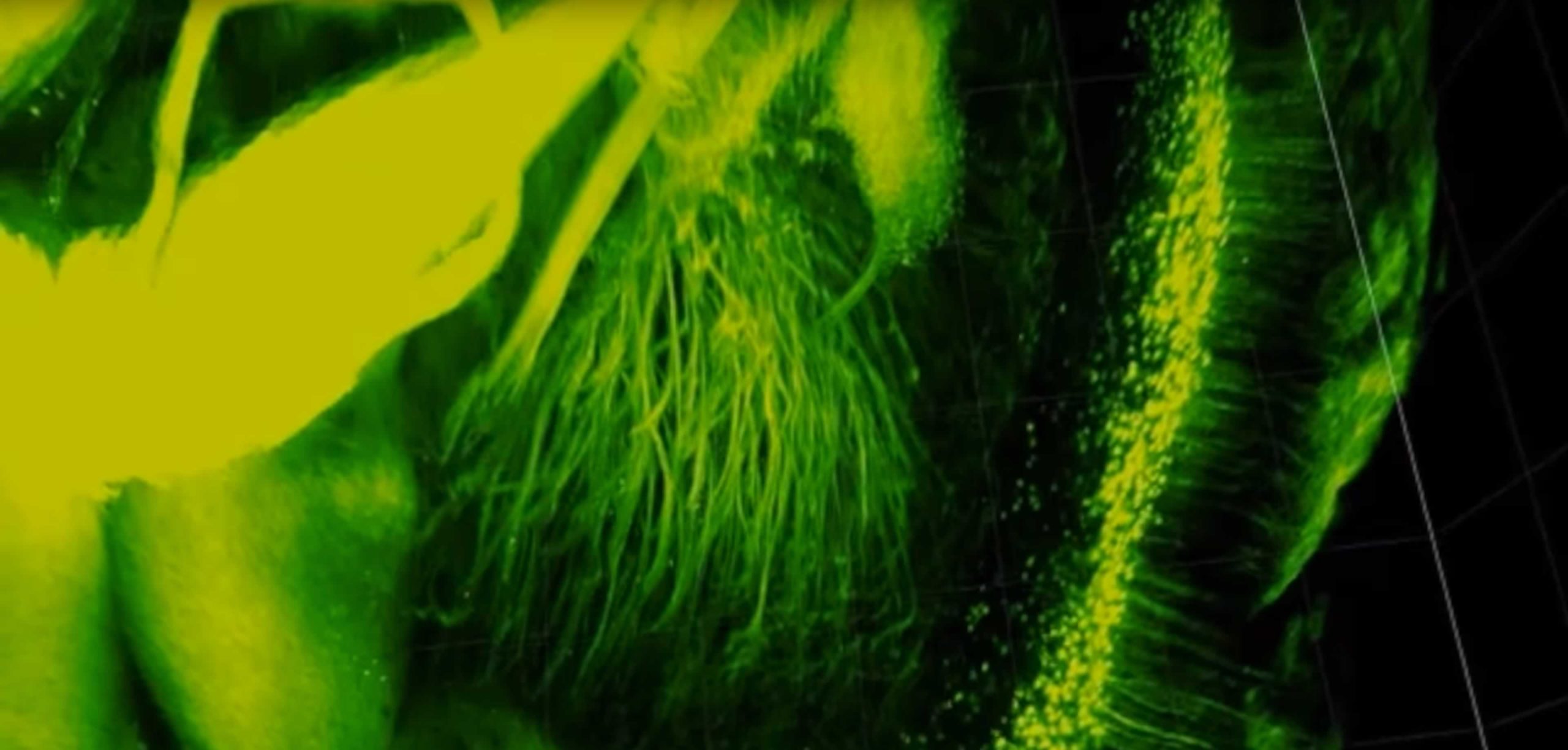
Single neurons stained and cleared with CLARITY
“CLARITY is powerful. It will enable researchers to study neurological diseases and disorders, focusing on diseased or damaged structures without losing a global perspective. That’s something we’ve never before been able to do in three dimensions.”
-Francis Collins, Director of the National Institutes of Health (NIH)

Next Generation Neurological Imaging of Mouse Cortex
3D Subcellular Brain Tissue Imaging
With nondestructive CLARITY tissue clearing, scientists can spatially identify the interaction of a variety of cellular phenotypes within the same tissue sample. Since the sample is embedded and remains intact within the cleared hydrogel matrix, the stained tissue has the potential to be imaged entirely in three dimensions. The tissue’s transparency enables 3D imaging of subcellular components such as DNA, RNA, and proteins for analysis of heterogeneous cellular interactions within the tissue microenvironment.
Learn more about ClearLight Bio
Unclouding the Brain
Previously, it has been difficult to see inside a brain. As an extremely lipid-rich tissue, a milky opaque tissue coats much of the brain, clouding the view. That makes light microsocopy of the brain largely ineffective. Without being able to see it’s hard to get a picture of the neural structure and circuitry. With CLARITY tissue clearing and 3D tissue analysis in 3D this fundamentally changes.
The Brain is Open for Discovery
New Era for Neuroscience
Subcellular Brain Tissue Imaging
Karl Deisseroth: "You can actually fly in and look around to see which connections are next to which other connections. Where you see groups of greenish yellow spheres -- those are individual neurons. And, you can see how they’re all arranged relative to each other, and the different colors -- the green, the red and the blue -- each of those are labels for a different kind of cell. And, our goal is to understand the system in its entirety, but also at high resolution, at the level of its wiring and the individual cells."
Drug Development for Brain Diseases Could Benefit from Subcellular Imaging
")
The brain c.1508-9 by Leonardo da Vinci (Vinci 1452-Amboise 1519)
Technology in science and medicine has evolved dramatically since the time of Renaissance man, Leonardo Da Vinci’s famous sketching of the human anatomy over 500 years ago. Imagine if he could witness subcellular brain tissue imaging.
Clinical therapies and medications have been developed to treat conditions including Alzheimer's disease, Parkinson's disease, Dementia, Epilepsy and Other Seizure Disorders, Stroke, Brain Cancer, and other Autoimmune and Neurodegenerative diseases. However, more drug discovery and development is needed. These diseases still regularly cut lives short and reduce human potential.
We believe the most effective medications have yet to be developed. At ClearLight Biotechnologies we are developing a platform to empower scientists and researchers to develop next generation cures. New psychiatric drugs, neurological drug development, and novel brain biomarkers are more likely to be targeted based on a better understanding at the molecular and physiological level.
Perhaps you have the curiosity, inventiveness, and resourcefulness of Leonardo Da Vinci. Are you a modern-day Renaissance woman or man who is building on the achievements of yesterday’s science heroes with the promises of today’s advanced technologies? Surely, the cures to all of the world’s diseases haven’t already been created.
Beautiful, Surreal Drawings of the Brain by the Father of Modern Neuroscience
"The Beautiful Brain" at NYU's Grey Art Gallery features the drawings of Santiago Ramón y Cajal (Spain, 1852–1934). Known as the father of modern neuroscience, Cajal is credited with discovering intricate functions of the brain long before the benefits of modern medical imaging.
The Father of Modern Neuroscience
Moving forward in time 400 years beyond da Vinci, Santiago Ramón y Cajal, a Spanish pathologist, specializing in neuroanatomy earned the title of The father of modern Neuroscience. His pioneering investigations of the microscopic structure of the brain and work on the structure of the nervous system earned him a Nobel Prize in Physiology or Medicine in 1906.
“Any man could, if he were so inclined, be the sculptor of his own brain.”
ClearLight Biotechnologies Scientific Advisors

Karl Deisseroth, MD, PhD Founder & Scientific Advisor
Dr. Karl Deisseroth, professor of bioengineering and psychiatry at Stanford University, founded ClearLight based on the CLARITY lipid-clearing technique he and colleagues developed at Stanford University. His scientific achievements have spawned additional research and methods that have revolutionized the study of the brain and have led to major advances in neuroscience and biomedical engineering.
“We need to take big risks and even blind leaps.”
See More Biology and Explore Further.
Get in touch with us when you want to see more biology and explore further. Your research deserves it.
Future patients deserve it.
