Tissue Clearing Applications
Lung Tissue & Lymph Nodes
Learn More About Tissue Clearing
- Tissue Clearing Methods Compared
- CLARITY Tissue Clearing Primer
- CLARITY Breast Cancer Study
- Moving Beyond Slices and Slides
Supporting Your Research Application
Tissue Clearing Lung Tissue
ClearLight has applied CLARITY tissue clearing, immunohistochemistry, and Tru3D image analysis to healthy lung tissue, lung carcinomas, and associated lymph nodes.
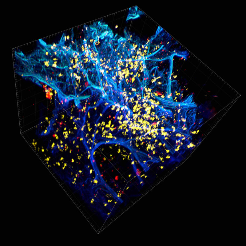
Video showing human non-small cell lung cancer adenosquamous tumor (NSCLC) associated metastatic lymph node: DAPI (blue), pan-cadherin (green), CD8 (yellow), α-SMA (red).
Tissue Cleared and Immunolabeled Human Metastatic Lymph Node
Image of human metastatic lymph node associated with a Stage 2A non-small cell lung cancer adenosquamous tumor. Pan-cadherin (green), a cell adhesion marker, was used to observe tumor cells, while CD8 (yellow) denoted the cytotoxic T cells, and alpha smooth muscle actin (α-SMA) shown in red, was used as a marker for stromal cells. DAPI, our nuclear counterstain is shown in blue.
Try tissue clearing on your own.
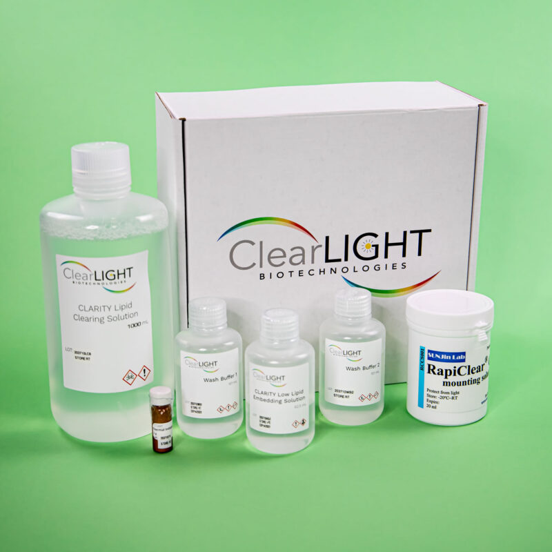
If you want to apply CLARITY to the tissues highlighted on this page, you will need to use the ClearLight LOW Lipid Tissue Clearing Kit.

Leverage our Internal R & D
When you work with us you gain the benefit of our internal experiments, assessments, and rigorous testing. In addition to performing our lab services: CLARITY Tissue Clearing, 3D IHC, and Tru3D Tissue Analysis, ClearLight scientists perform internal Research and Development to advance our understanding and applications of CLARITY to other tissue types and downstream applications. Researchers who work with us benefit from this exploratory work. The ClearLight “lab basement” is full of failed or pivoted experiments that helped inform our path to arrive at what does work. Work with us to avoid making the same mistakes that we have made or going down the blind alleys that we’ve pivoted away from.
“If you don’t want to spend the time figuring it out, we can help. We work hard to make this look easy.” - Dr. Sharla White, VP of Research and Development
Ready to Discuss Your Research Question? Meet with our Application Scientist.


