CLARITY Tissue Clearing
Creating See-through Tissues
How we prepare tissues for antibody labeling and IHC Multiplexing
CLARITY Tissue Clearing Navigator
- Limitations of Imaging
- Purpose of Tissue Clearing
- Why CLARITY for Tissue Clearing?
- CLARITY Tissue Clearing Overview
- CLARITY Tissue Clearing Step-by-Step
- CLARITY 101 Video
- CLARITY Advantages
- Passive vs Active CLARITY
- CLARITY FAQs
- ClearLight Advantage for CLARITY
- Diagnostic and Clinical Applications
- Three Ways to Leverage ClearLight
- Join Creating Cures Community

Tissue Clearing Primer
CLARITY is an acronym for Clear Lipid-exchanged Acrylamide-hybridized Rigid Imaging / Immunostaining / in situ-hybridization-compatible Tissue hYdrogel). Discussions about CLARITY tissue clearing inevitably involve imaging. The two go hand-in-hand. Tissue clearing is what you need to do in order to see beyond the tissue surface at a cellular and subcellular level. It is akin to being able to see in the darkness using night vision goggles or using sunglasses to be able to clearly see the landscape in the presence of bright sunlight. When researchers and scientists want to see inside of a brain, or tumor or study diseased or healthy tissue they first will want to have that tissue cleared of the things that occlude the view.
Imaging Tissues, Bodies and Biological Systems

Most tissue is opaque, meaning that visible light cannot travel through it. For example, you can’t see through your hand. If you hold your hand up to a light source, it will block the light and cast a shadow. If you fall and injure your hand, your doctor can use an x-ray machine to see if you may have broken a bone.

Unlike visible light, x-rays pass easily through the soft tissue of your hand, like skin, muscle and tendons, but they scatter when they encounter dense material such as bone. Because of this property, x-rays are ideal for imaging broken bones and discovering tooth decay.

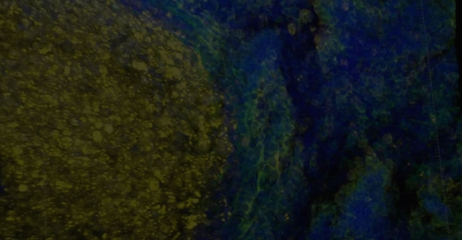
, pan-cadherin (green), CD8 (yellow), α-SMA (red)")
Limitations of Imaging
To study structures in soft tissue—especially very small, delicate structures, like neurons in a brain, or cancerous lesions in breast tissue, scientists need a way to see beyond the tissue surface.
Historically, this meant putting thin slices of tissue onto glass slides and then staining and examining the slides one by one under a microscope. They could only guess how the tiny tissue structures actually looked and functioned in the whole sample before being dissected and destroyed in the process of analysis.
These techniques are over 100 years old, and yet still represent the current gold standard. Scientists and researchers still primarily rely on the analysis of 2D thin section FFPE (~5-10 micron) samples. Using these methods, spatial and morphological analysis of the tissue microenvironment is constrained. Additional limitations include the tedious and labor intensive processing of hundreds of slides. Therefore, researchers using the current gold standard rely on a small sampling of the tissue in attempting to understand complex biological processes. This is the problem with 2D FFPE.
Tissue clearing exists to improve imaging.
Although rudimentary types of tissue clearing have been around since the early 20th century, they were mostly useful for large samples. Small and subcellular details were destroyed in these early methods. In the last 5 - 10 years, modern tissue clearing techniques, especially those that can be combined with immunohistochemical staining techniques, are revolutionizing how we study subcellular tissue morphology.
Enter the Microenvironment
Tissue clearing enables researchers to study cancer, neurological diseases and many other disorders that require small-scale, tissue-level imaging, allowing scientists to focus on diseased or damaged structures without losing a global perspective. That’s something we’ve never before been able to do in three dimensions before recent technological advancements. We can now enter the microenvironment.
You need to image tissue in order to understand its structure and study its components. This is necessary for basic research and more urgently necessary in medicine where a correct diagnosis precedes successful treatment. The more scientists and medical practitioners can understand the impact of disease on the body, the greater the chances they can create the treatments and cures that will alleviate suffering and extend human life.
Why CLARITY for Tissue Clearing?
CLARITY Tissue Clearing Process
CLARITY was born from the need to overcome the opacity of lipids in brain tissue, which caused light to scatter during microscopic visualization of neurons, thereby obscuring image quality. CLARITY allows a tissue sample to be rendered optically clear, retain its three-dimensional integrity and allows for tissue staining and re-interrogation multiple times. Though originally conceived to make brains transparent to light, CLARITY tissue clearing methods have now been used on multiple human, mouse and rat tumors, organs, bone and most other intact biological tissues, in addition to the brain.
CLARITY Tissue Clearing Genesis
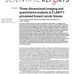
The CLARITY method was originally developed at the Karl Deisseroth Lab at Stanford University. It was first made public in a paper published in Nature in 2013. [ K Chung, J Wallace, S-Y Kim, S Kalyanasundaram, AS Andalman, TJ Davidson, JJ Mirzabekov, KA Zalocusky, J Mattis, AK Denisin, S Pak, H Bernstein, C Ramakrishnan, L Grosenick, V Gradinaru, and K Deisseroth. Structural and molecular interrogation of intact biological systems. Nature (2013) 497: 332-337. ]
Creating See-through Tissues
CLARITY Tissue Clearing Overview
In the CLARITY method, a tissue sample is first embedded in a hydrogel. This hydrogel provides a three-dimensional matrix that supports the tissue sample and retains the structural integrity and spatial orientation of the critical tissue components. The non-critical elements such as lipids are removed using detergent solutions. Light can then penetrate the tissue sample rendering it “see-through” or transparent.

Specific steps in the ClearLight CLARITY Tissue Clearing Process

Hydrogel Matrix
The first step in the tissue processing workflow is to place the fixed mouse, rat, or human tissue sample in a solution of hydrogel monomers and cross-linkers. For detailed explanations refer to Hydrogel-Tissue Chemistry.

Biomolecule Functionalization
The monomers and cross-linkers diffuse into the tissue’s cells and bind to biomolecules such as proteins and nucleic acids but not to light-scattering lipids.

Polymerization
The hydrogel is thermally treated and the monomers polymerize into stable mesh that locks proteins and biomolecules in place.

Lipid Clearing
A detergent is used to remove lipids and other unbound molecules from the tissue. The proteins, nucleic acids and other bound biomolecules remain embedded with the hydrogel mesh.

Immunostaining
If desired, antibody-based immunostaining or labeling for many nucleic acids (RNA/DNA) can be used in a multiplex panel to highlight specific structures in the tissue cleared sample. Learn more about immunohistochemistry (3D IHC).

Image Analysis
The tissue processing workflow moves from molecular labeling to imaging. The stained tissue is placed in a refractive index-matching mounting solution for imaging with a confocal or light sheet microscope or another imaging platform.

CLARITY Advantages
Benefits of using CLARITY in Tissue Processing Workflow
- Compatible with previously frozen, fresh, formalin-fixed, and FFPE embedded tissue
- Compatible with traditional pathology workflow methods
- Non-destructive, fewer samples, allowing recapitulation of spatial heterogeneity without laborious sectioning and registering
- No dehydration/rehydration steps involved as with FFPE samples, or other tissue clearing technologies
- Compatible with standard nucleic acid and antibody interrogation techniques
- Allows multiple interrogations of a single sample to increase 3D biomarker information
- Samples can be stored for future biomarker analysis
- Imaging compatibilities include: Confocal, Light Sheet, SPIM
Passive CLARITY vs Active CLARITY (CRYSTAL)
One of the disadvantages of the CLARITY method is the time it can take to fully clear a sample. To deal with this issue researchers have invented active CLARITY methods, most notably the CRYSTAL protocol that dramatically reduces the overall timeframes involved.
With CRYSTAL, detergent streams are directed at the tissue. These streams act like a pressure washer for the tissue sample, actively targeting particular areas instead of waiting for the passive diffusion.
To visualize this effect we can compare it to a situation we can all relate to. Imagine you are washing a dirty pan that has food stuck on the bottom. There are two basic approaches to cleaning the pan. You can let the pan soak for hours until the hardened food debris softens, or you can vigorously scrub and get the pan cleaned faster by putting in physical effort. Letting the pan soak is similar to passive CLARITY, while actively scrubbing the pan clean is like using CRYSTAL along with CLARITY.
While CRYSTAL technology is relatively new, we have a system currently in development that will help automate this process, and help make CLARITY practical for clinical settings, as well as research labs.

Services
We currently perform non-destructive tissue processing to facilitate pre-clinical research studies.
See Sample Requirements on the Services page. We may support situations not shown. Start a conversation with us using the Contact Us form, email, call (408) 329-7769 or chat with us online.
ClearLight provides CLARITY Tissue Clearing Kits to researchers who wish to perform lipid clearing of soft tissues on their own using the CLARITY method. Visit the online store to learn more about these products. Researchers can also engage lab services for CLARITY Tissue Clearing, 3D Immunohistochemistry and Tru3D Tissue Analysis. Visit Services to learn more.
Technology
Active or passive CLARITY tissue clearing is a very powerful and useful method, but lipid clearing and staining times take longer time than optimal for use in future clinical and diagnostic settings. CRYSTAL technology is a significant improvement to both the active and passive CLARITY tissue clearing methods by aiming detergent streams at specific areas of the tissue sample to be cleared. This can speed up the clearing process significantly, increasing the diagnostic relevance and usefulness of this technology in a clinical setting.
No, the CLARITY method was originally developed at the Karl Deisseroth Lab at Stanford University Medical Center. Dr. Deisseroth is a founder and scientific advisor to ClearLight Biotechnologies. However, ClearLight is automating the method so that it can be utilized by researchers and eventually clinicians across the world.
Why ClearLight for CLARITY?
ClearLight Scientists are Experts at CLARITY
Although CLARITY protocols have been published for several years, CLARITY tissue clearing can be difficult to get right. Many scientists and researchers who could benefit from using CLARITY do not have the experience or the time to master using these methods in the lab.
Instruments to automate the CLARITY tissue clearing process have fallen short of expectations. First, the cost of the machines is prohibitive, especially for researchers working off of grants. Second, many of these automated systems, in a working lab environment, fail to perform as designed. The quality of the clearing and staining work fails to deliver the full potential the CLARITY method has to offer.
In addition to the robust science behind CLARITY, there is an art to getting the best results using CLARITY tissue clearing. Our scientific team are experts in using CLARITY.
CLARITY started with our founder, Karl Deisseroth. CLARITY tissue clearing is foundational to ClearLight Biotechnologies. This is unlike other companies which sell reagents, tissue clearing kits, automated clearing instruments, etc.
ClearLight Biotechnologies is a contract research partner who can provide expert CLARITY tissue clearing, immunohistochemistry, and 3D imaging so researchers and scientists can see more biology.
ClearLight Biotechnologies has an exclusive, worldwide license agreement with Stanford University for development and commercialization of CLARITY for diagnostic and clinical applications.
CLARITY for Diagnostic and Clinical Applications
Imagine being able to peer inside the tumor microenvironment. Imaging fueling the power of your pre-clinical research. Imagine how this can empower better clinical diagnosis, prognosis, and predictive applications in the future. Imagine such capable instruments being deployed in every research and pathology laboratory around the world. Imagine immuno-oncology empowered with Tru3D imaging and tissue analysis and. This is the future we are working to make possible. We invite you to collaborate with us.
CLARITY Tissue Clearing Infographic
Three Ways to Leverage ClearLight
for your Research

ClearLight Lab Services
ClearLight Biotechnologies lab services include tissue clearing using the CLARITY method, 3D immunostaining, 3D imaging (multiplexed fluorescence), and Tru3D tissue analysis.
")
Tissue Clearing Kits
CLARITY Tissue Clearing Kits are for researchers who wish to perform lipid clearing of soft tissues on their own. Research Use Only Kits available in both High Lipid and Low Lipid versions.

Pursue Pilot Study
Our Implantable Device Case Study shows how we helped a biomedical innovator design a better implantable device for drug delivery. Bring us your burning research question.
