FFPE vs 3D IHC
Compare FFPE to CLARITY + Tru3D
A comparison of 2D tissue processing and 3D Tissue analysis
The Problem with 2D FFPE
Current gold standard techniques are over 100 years old and rely on the analysis of 2D thin section FFPE (~5-10 micron). Spatial and morphological analysis of the tissue microenvironment is highly limited as well as the limitations associated with processing hundreds of slides. Therefore, researchers rely on a small sampling of the tissue to understand complex biological processes.
Standard General Workflow - FFPE IHC Imaging

FFPE Block

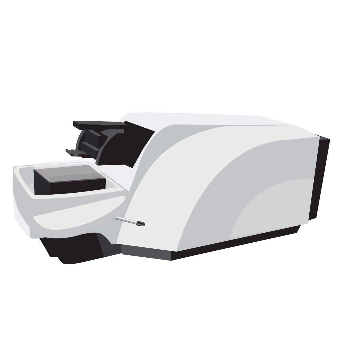
Microtome - FFPE Sectioning

5 Micron section FFPE Tissue

Confocal Microscope - Immunofluorescence
Whole Slide or Field of View Imaging
Standard Pathology Measurements
Maximum tissue size limitations:
- Histology tissue cassette (standard size)
- Block face: 30 x 25 mm (3 x 2.5 cm)
- A margin of 2 mm of paraffin around all sides of tissue is preferred for proper support during cutting
- Depth: 4 mm (0.4 cm)
- Embedded tissue must be 3 mm (0.3 cm) thick or less
- A paraffin margin preferred in order to section entire sample.
- Estimated workable dimensions for maximum tissue embedding
- 26 x 21 x 0.3 mm
- Length x width x depth/thickness
- Block face: 30 x 25 mm (3 x 2.5 cm)
- Microscope slide (workable space – standard size): 40 x 22 mm
Volumetric Comparison
FFPE
- Estimated maximum size of tissue onto slide: 25 x 20 mm (2.5 x 2 cm)
- Estimated maximum depth/height of entire FFPE tissue block without additional sectioning: 3 mm
- Estimated unsectioned maximum volume: 1500 mm3 (1.5 cm3)
- FFPE tissue section sample (maximum XY dimensions, 5 µm section): 25 mm x 20 mm x 0.005 mm thickness
- Volume: 2.5 mm3 (0.25 cm3)
Tru3D and CLARITY
- Standard entire ClearLight Biotechnologies tissue sample dimensions: 5 mm diameter (radius: 2.5 mm) x 1 mm thickness
- Volume: 19.6 mm3 (1.96 cm3)
- Standard ClearLight Biotechnologies tissue sample (6 FOVs) dimensions: 0.454 x 0.454 x 0.5 mm
- Volume: 0.106 mm3 (0.0106 cm3) per FOV
- Total volume (6 FOVs): 0.618 mm3 (0.0618 cm3)
- Imaging parameters: 25x objective
Sample Processing Workflow - ClearLight Biotechnologies
The tissue/tumor is shipped to ClearLight Biotechnologies. We slice it with our vibratome into 1 mm thick slices.
That slice may or may not be further sectioned into a 5 mm dia. x 1 mm piece.

Tumor / Tissue
Vibratome - 1 mm sections

Tumor sliced (1 mm ea.)

5 mm x 1 mm Tissue Sample
Why We Use 6 Fields of View

5 Micron section FFPE Tissue
Volume: 0.618 mm3
Tissue Dimensions: 11x11 mm (equiv.)

5mm dia. X 1 Field of View
Volume: 0.618 mm3
(Current)
A comparable 5 µm FFPE section to our current ClearLight Biotechnologies 6 FOVs would have the XY dimensions of 11 x 11 mm or essentially half of the maximum area; however, without the ability to look deeper into the section, a potential lesion could still be missed. While we are not currently imaging the entire 5 mm dia. x 1 mm ClearLight Biotechnologies sample, we are scanning the entire sample, without image capture, to identify any potential lesion. We then perform imaging in the proper FOV that contains the lesion, thereby not missing it during selection.

Compare 2D FFPE with CLARITY + Tru3D™
Comparison* of volumetric data - 1 field of view

Imaged Sample Type
* assumes same imaging settings
ClearLight Produces More Volumetric Data
How does the ClearLight Biotechnologies 6 FOVs assessment compare to a clinical biopsy sample? For example, an invasive vacuum-assisted-device needle core biopsy provides a core biopsy with a diameter ~2.4 mm. The resulting volume from an FFPE section would be 0.025 mm3. If the entire biopsy was scanned, the current ClearLight Biotechnologies approach utilizing 6 FOVs would still provide ~25-fold more volumetric data. Theoretically, even imaging 10 core biopsies from the patient tumor would still provide less volumetric data than one ClearLight FOV image assessment.
CLARITY IHC Imaging FOV Workflow

5 mm dia. x 1 mm Whole Tissue Volume

Tissue Sample Cleared and Being Stained via IHC

Confocal Microscope - Immunofluorescence
Field of View Imaging
Tissue Sample Comparison
2D FFPE vs. CLARITY Tru3D
Gold Standard FFPE
5 μm Section - FFPE Tissue
CLARITY Processed Tissue

500-1000 μm Section - CLARITY Tissue
Imaging Comparison
2D FFPE vs. CLARITY Tru3D
Overcome limitations of 2D imaging which is not representative of the heterogeneous biopsy or tissue microenvironment. See videos to compare 2D FFPE with CLARITY + Tru3D.
3D Image Analysis in 2D

Gold Standard Formalin-Fixed Paraffin Embedded (FFPE)
2D technology

Processed by CLARITY technology and analyzed using proprietary AI-based software on 3D volumes of tissue
