Tissue Clearing Applications
Clearing Brain Tissue
Learn More About Tissue Clearing
- Tissue Clearing Methods Compared
- CLARITY Tissue Clearing Primer
- CLARITY Breast Cancer Study
- Moving Beyond Slices and Slides
Supporting Your Research Application

Cleared and Immunolabeled Brain Tissue

Using CLARITY to Remove Lipids in Brain Tissue
With the lipids removed from brain tissue, scientists can add molecular markers to highlight certain features. Then, normal and diseased brains can be compared. New understanding could bring new insights to create the next generation of therapies and cures.
Passively cleared and immunostained normal mouse brain (5 mm dia. x 1 mm thick) that has been stained with DAPI (blue) to identify individual cells, Lectin-DyLight 594 (red) to identify vasculature, and Histone H3-Alexa Fluor 647 (green), a chromatin marker.
CLARITY Inventor, Karl Deisseroth, Talks about CLARITY Applied to Brain Tissue
Try tissue clearing on your own.
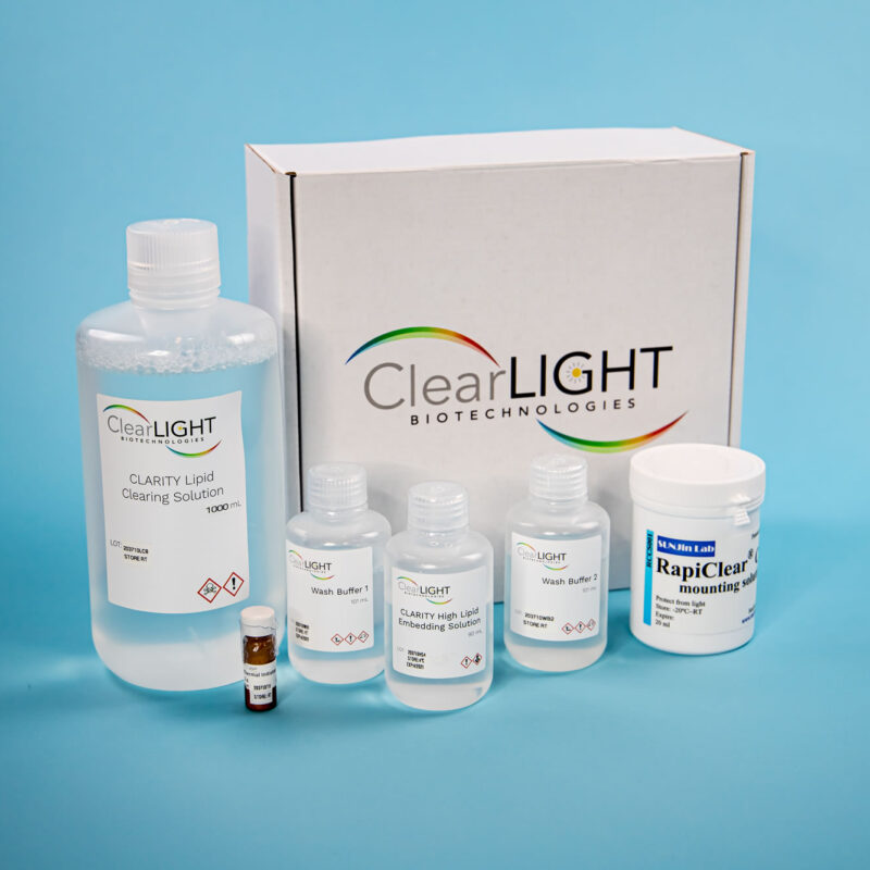
If you want to apply CLARITY to the tissues highlighted on this page, you will need to use the ClearLight HIGH Lipid Tissue Clearing Kit.

Karl Deisseroth, MD, PhD Founder & Scientific Advisor
Scientific Advisor
Dr. Karl Deisseroth, professor of bioengineering and psychiatry at Stanford University, founded ClearLight based on the CLARITY lipid-clearing technique he and colleagues developed at Stanford University.
“We need to take big risks and even blind leaps.”
You May Also Be Interested In:

Leverage our Internal R & D
When you work with us you gain the benefit of our internal experiments, assessments, and rigorous testing. In addition to performing our lab services: CLARITY Tissue Clearing, 3D IHC, and Tru3D Tissue Analysis, ClearLight scientists perform internal Research and Development to advance our understanding and applications of CLARITY to other tissue types and downstream applications. Researchers who work with us benefit from this exploratory work. The ClearLight “lab basement” is full of failed or pivoted experiments that helped inform our path to arrive at what does work. Work with us to avoid making the same mistakes that we have made or going down the blind alleys that we’ve pivoted away from.
“If you don’t want to spend the time figuring it out, we can help. We work hard to make this look easy.” - Dr. Sharla White, VP of Research and Development
Ready to Discuss Your Research Question? Meet with our Application Scientist.