Press
ClearLight Media Resources
Logos, images, videos, collateral and more.
Fast Facts
- Founded in 2015 by Karl Deisseroth MD, PhD
- Early-stage private company backed by Wiegers and affiliates, Pivotal and KESA Partners
- Experts at CLARITY Tissue Processing, thick tissue antibody staining, and Tru3D tissue analysis
- Provides research use only lab services to pharmaceutical, biotechnology, and academic researchers
- Developing next-generation tissue processing and image analysis platform to facilitate 3D analysis of preclinical and clinical models of disease
Company Overview
ClearLight Biotechnologies provides contract research and development services to pharmaceutical companies who seek to improve the efficacy of potential drug candidates. The company provides pharmaceutical, biotechnology, and academic researchers with CLARITY Tissue Clearing, 3D immunostaining, and Tru3D image analysis services. The company is developing an automated instrumentation platform based on the CLARITY lipid-clearing technique developed by Dr. Deisseroth and colleagues at Stanford University.
ClearLight Press Resources
Frequently used press resources including downloadable images, video, fast facts, logos, and more.
Image Usage Policy
While we don’t require prior approval for use of images and videos we do ask that you courtesy inform us of where and when you plan to use them. Use the embed codes or include the credit line on all photos and videos used, “Courtesy of ClearLight Biotechnologies.” Please use the embed link or include this credit on the page you use any of these images or videos.
Brand and Style
Logos, usage, and more.
Our Team
Dr. Sharla White
Vice President of Research and Development
- Expert in CLARITY across different tissues
- Diverse background brings perspective
- Experience with various pilots and feasibility studies
- Research focus areas:
- cancer biology
- immunotherapeutics
- cancer immunology
- tissue clearing technologies
- cardiovascular diseases
- vascular biology
- Inflammation


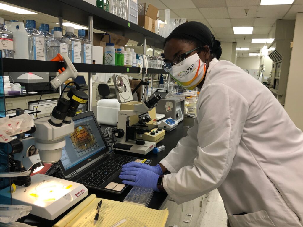
Press Resources - Images and Videos
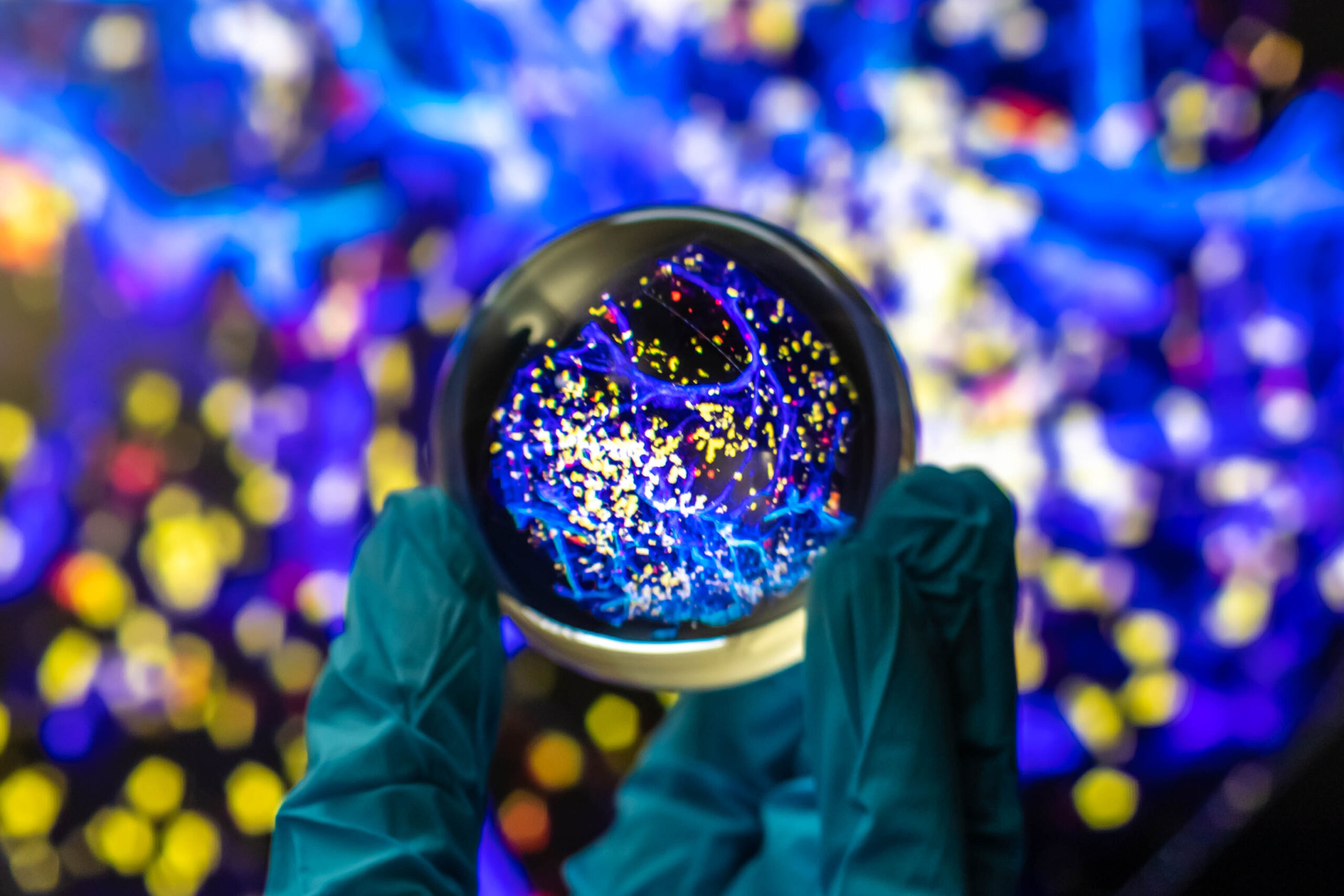
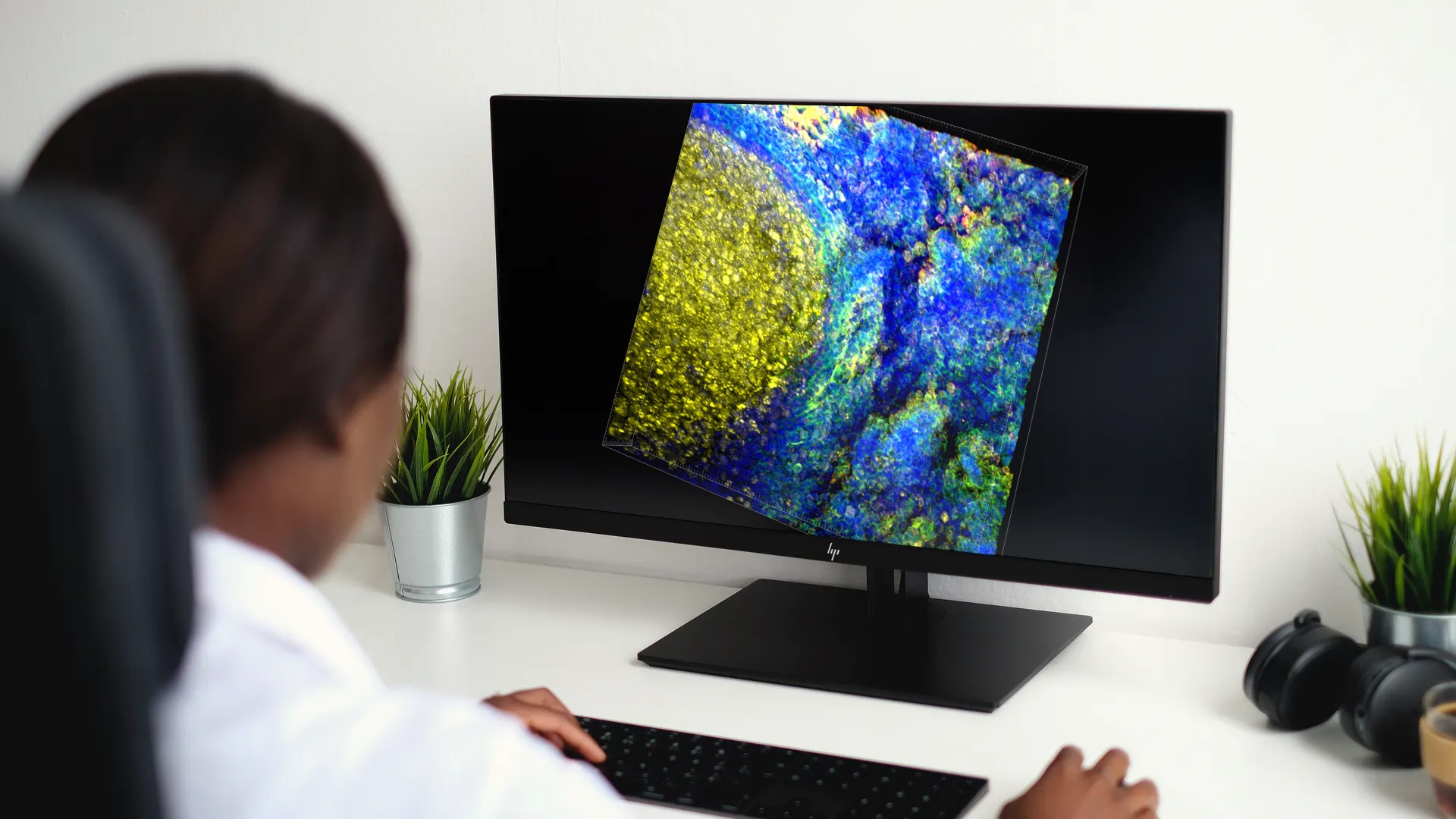
Image and video examples of immunostained tissues after CLARITY Tissue Clearing. #SeeMoreBiology
Selected Press Resources Images
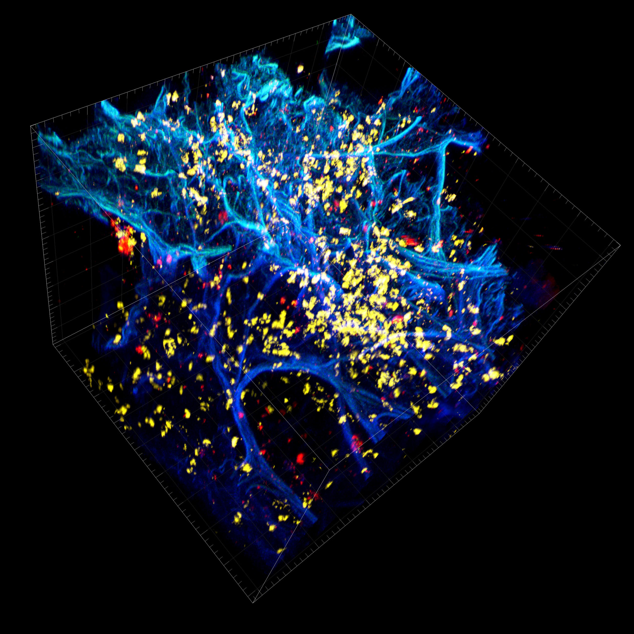
Lung Carcinoma
DAPI (blue), pan-cadherin (green), CD8 (yellow), α-SMA (red)
, pan-cadherin (green), CD8 (yellow), α-SMA (red)")
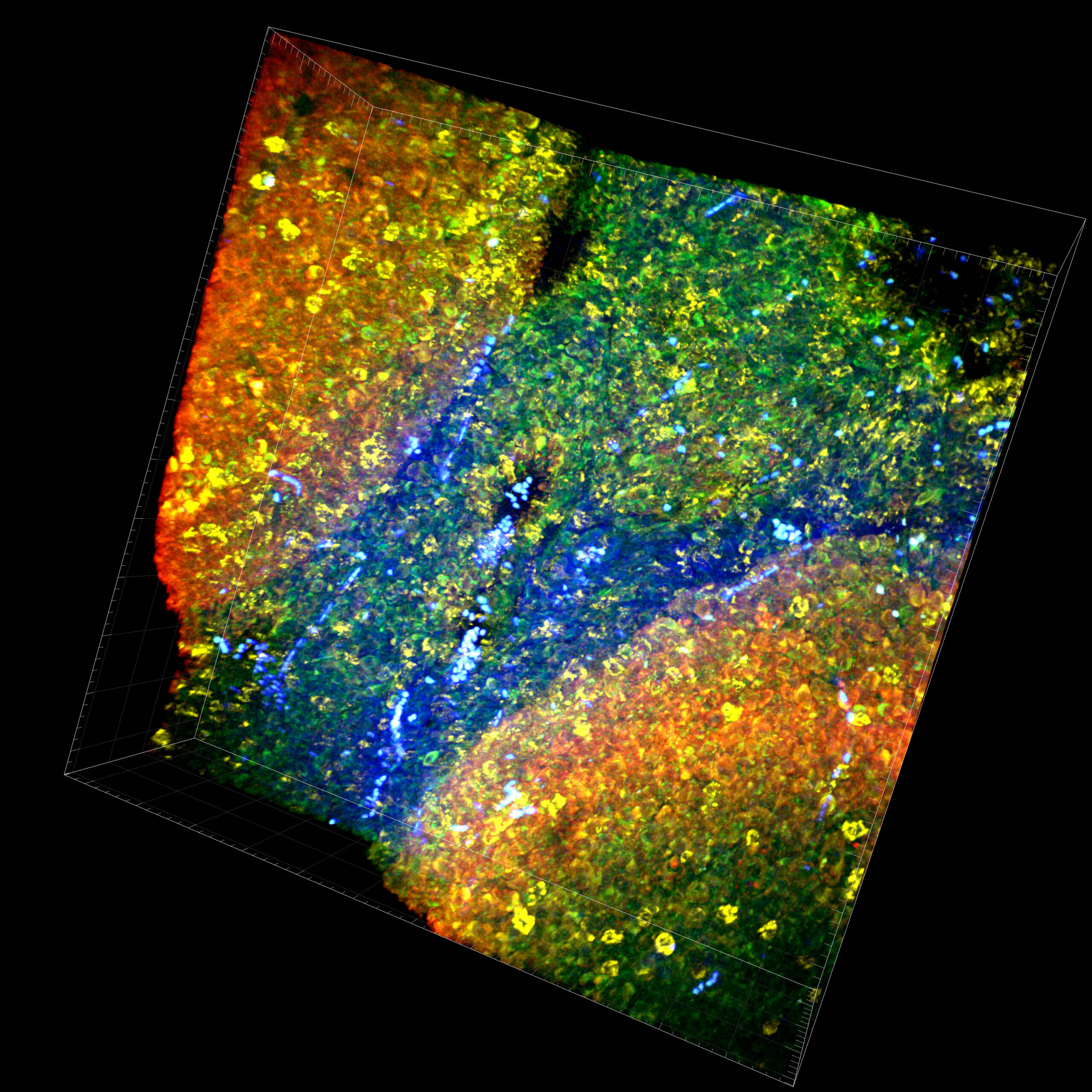
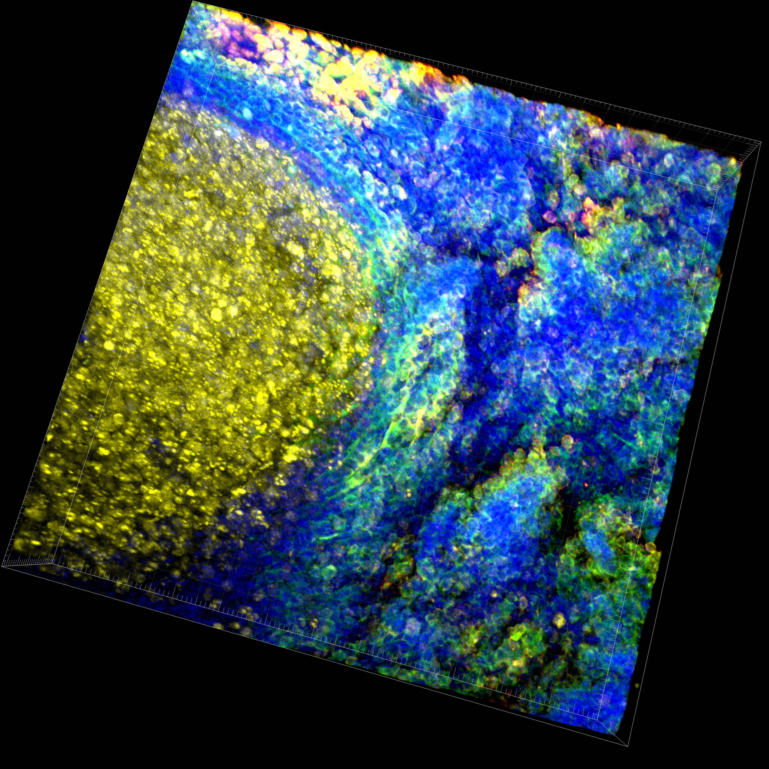
Human Tonsil
DAPI (blue), CD8 (yellow), pan-Cytokeratin (green), PD-L1 (red)
, CD8 (yellow), pan-Cytokeratin (green), PD-L1 (red)")
, Ki67 (yellow), pan-Cytokeratin (green), CD3 (red)")


Selected Press Resources Videos - Examples of 3D IHC

Video Loop 1:
A passively cleared and immunostained normal mouse brain (5 mm dia. x 1 mm thick) that has been stained with DAPI to identify individual cells, Lectin-DyLight 594 to identify vasculature, and Histone H3-Alexa Fluor 647, a chromatin marker.
, CD31 and DAPI")
Video Loop 2:
A vasculature-rich region of an inflamed human tonsil tissue that has been immunostained with CD4 to denote immune cells, such as monocytes, T helper cells, dendritic cells, and macrophages. Both alpha smooth muscle actin (α-SMA) and CD31, primarily an endothelial cell marker, were used to observe possible vasculature with some signal co-localization observed in certain vessels. Finally, the nuclear stain, DAPI, is shown to demonstrate all cells present in the tonsil tissue.
, CD163, CD68, and DAPI")
Video Loop 3:
An image of a Hep55 syngeneic mouse tumor, used to model hepatocellular carcinoma, that has been immunostained for pan-cytokeratin (pan-CK), CD163, CD68, and DAPI, a nuclear cell DNA marker. Pan-CK can be used to identify epithelial cells and denote tumor cells, while CD163 and CD68 can identify immune cells and macrophage-linage cells.

Video Loop 4:
An inflamed hyperplastic tonsil tissue immunostained to identify epithelial cells (pan-cytokeratin), proliferating cells (Ki-67), and T cells (CD3). DAPI, the nuclear counterstain, marks all cells present in the tissue.

Video Loop 5:
An inflamed human tonsil tissue stained with DAPI to clearly identified the individual cell nuclei present within the tissue. Both alpha smooth muscle actin (α-SMA) and CD31 were used to observe possible vasculature with some signal co-localization observed in certain vessels. CD45RO was used a marker to aid in the identification of regulatory T cells (Tregs).

Video Loop 6:
An inflamed human tonsil tissue stained with DAPI to clearly identified the individual cell nuclei present within the tissue. Both alpha smooth muscle actin (α-SMA) and CD31 were used to observe possible vasculature with some signal co-localization observed in certain vessels. CD20 is utilized as a pan-B cell marker and used identify B cells, and in rare cases can be found on some T cells.


How Can We Help You?
Should you require something more than the press resources on this page, we will be happy to assist you. Please direct any questions to JUG Marketing, at (657) 221-7128. If you’d like to be contacted directly, please fill out the form below and include specific media inquiry details.